*In this study, its tried to be understood that how retinotopic surface maps are changed and are they improved to define visual areas.
*Two different functional scans are compared, one is Single Shot EPI with ring stimulation given to subject the other is the Multiplexed EPI sequence which acquires twice many images in the same time.
*Check for sequence and stimuli in detail:
http://visionumram.blogspot.com/p/retinotopy.html
http://visionumram.blogspot.com/p/mepi-epi-comparison-by-glm-and-ica.html
*Mosaic image acquired from Multiplexed EPI
image has geometrically distorted and artefact at borders of the head.
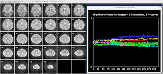
*Motion Correction of the Multiplexed EPI
*Registration
Distortion around visual cortex (sagittal) is visible,registration at center is good.
*Time course of the activation from arbitrary ROI (EPI(upper),M_EPI(lower));
as the sampling rate increased, and BOLD amplitude increased however there is an high frequency noise source introduced,so advantages should be observed well.
*Surface Maps of EPI vs M_EPI
Single Shot EPI with Expanding Ring Stimulation
Multiplexed EPI with Expanding Ring Stimulation
*Comments&Conclusion
-Ring is quite visible in M_EPI and spatial specificity is high.
-V1 dorsal border is shown on ring stimulation,it can be said that V1 border at single shot EPI is a little shifted in M_EPI but still perpendicular to ring.
-Spatial specifity is increased in M_EPI ring shown in more gradual color coding, but it needs further experimentation to say that borders are more distinct,and the error should be well studied because there are both additional perturbations at time courses and spatial maps due to geometric distortion at borders.
-Slice prescription with visual cortex at center may be more useful to avoid geometric distortion.
-Slice prescription with visual cortex at center may be more useful to avoid geometric distortion.


